It has been said that a picture is worth a thousand words. Under the lens of a microscope or novel imaging techniques, that photo can actually tell us much more, draw us closer to its subject and expose a world unknown.
That is the concept behind Science Exposed, the annual research photo competition from the Natural Sciences and Engineering Research Council of Canada (NSERC). At stake are three juried prizes and one People’s Choice Award, voted on by the public, with winners announced in the fall.
Science Exposed welcomes entries of scientific research in all fields of study to foster scientific curiosity while simultaneously building a database of Canadian research images.
This year, two members of the University of Guelph community are among the 20 finalists: master’s student Gabriella Willan from the Department of Biomedical Sciences at the Ontario Veterinary College; and PhD candidate Angie Homez from the Department of Food Science in the Ontario Agricultural College.
View the entries from the 20 finalists and vote here.
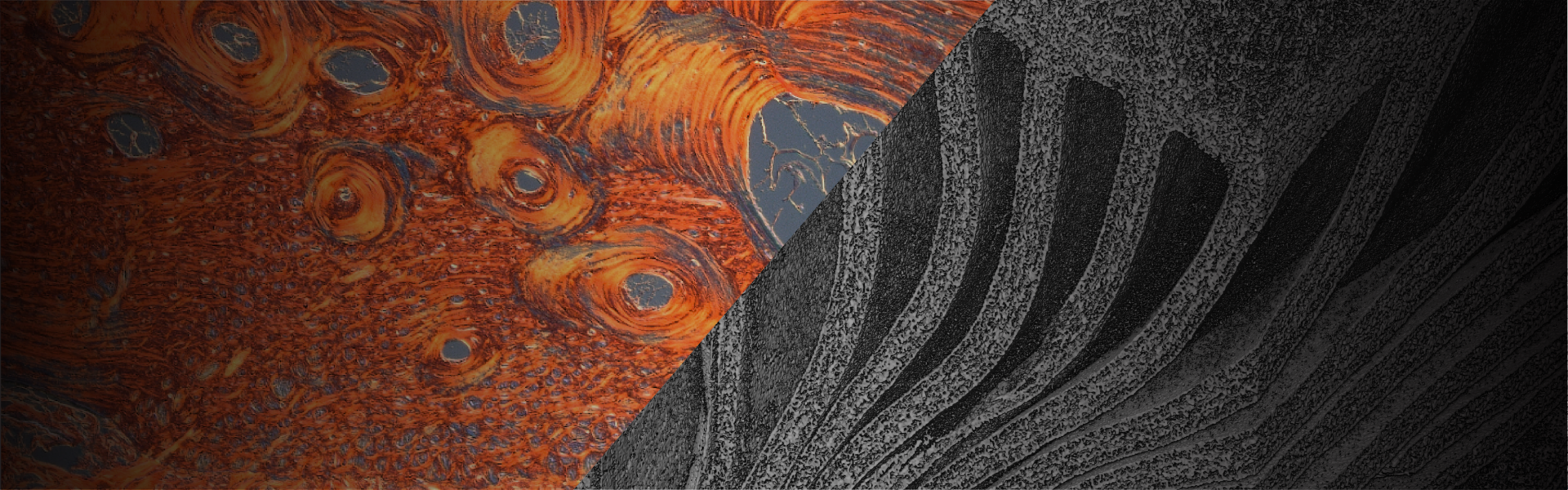
Biological armour: Rethinking the skeleton in the skin
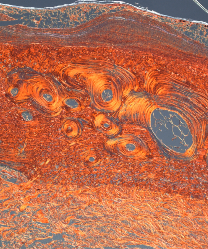
It was her colleagues in Dr. Matt Vickaryous’ lab in the Department of Biomedical Sciences that were first amazed by the image Gabriella Willan took of the layers of a lizard’s skin.
Many reptiles and amphibians have small, bony deposits in the skin that form an internal chain mail like meshwork, called osteoderms. The ancient structure, often thought of in relation to armadillos, has only recently started to be studied.
“It turns out lizards are doing something similar,” Willan says. “They’re putting bone in the layers of their skin. A lot of people think it is a protective mechanism.”
Projects like hers are rare and Willan had to develop histological techniques to be able to view such small slices of bone. Previous research determined the size and shape of osteoderms vary among lizards, but Willan says no one had taken a detailed look at tissue composition or how it might vary among species.
“My main question going into this was, what are lizards building these out of and are they all building them the same?”
The Australian shingleback lizard in Willan’s image showed her there was no clear correlation among species. The patterns in the image depict collagen fibre organization. The swirling patterns are organized bone. Above that is woven bone and on top of that is capping tissue. The holes in the image are marrow cavities.
“In a nutshell, that’s what you’re seeing,” she says.
Willan envisions this research reaching far beyond lizard skin. There are many avenues to explore, she says. For example, biomimetics. “Can we make better protective gear for sports? Can we make better helmets with better cushioning?”
“Science is relevant to everyone,” Willan says, “and everyone should get a chance to see what Canadian researchers are up to.”
Channels to food sustainability: the insight of mushrooms
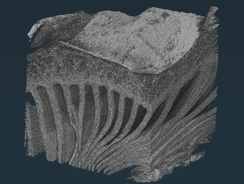
In Dr. Maria Corradini’s lab in the Department of Food Science, PhD candidates like Angie Homez are conducting research focused on the engineering aspects of food to better understand food spoilage.
Homez’s 3D image shows the microstructure of an oyster mushroom, created with micro-computer tomography. “One thing we’re trying to see is how the pores and structure of the mushroom break down,” Corradini explains.
“Mushrooms are quite fascinating,” Homez says. “I think that also drove my curiosity toward them, that they are in this unique location in the tree of life. They’re not plants or animals. They’re their own thing.”
The ability to see and analyze such tiny details allows researchers to measure porosity (the volume of channels or empty spaces) and tortuosity (the curvature of these channels). This provides clues to the stability of a food product. Corradini says they work with mushrooms because they’re sustainable and can be locally produced.
The photo’s level of resolution was made possible by using the Canadian Light Source’s synchrotron and leveraging the expertise of Jarvis Stobbs. This device produces powerful light beams to examine molecular and atomic details of materials. It is the only one in Canada and one of only a handful in North America.
The image of the oyster mushroom represents more than just the mushroom itself, Corradini says. It helps people understand the microstructure of food items and inform strategies that extend shelf life and reduce food waste.
“I think it’s a really nice way to get exposure and draw attention to what we’re doing,” Homez says. “It’s exciting, it’s challenging, but it’s also so rewarding when you see results like these.”