
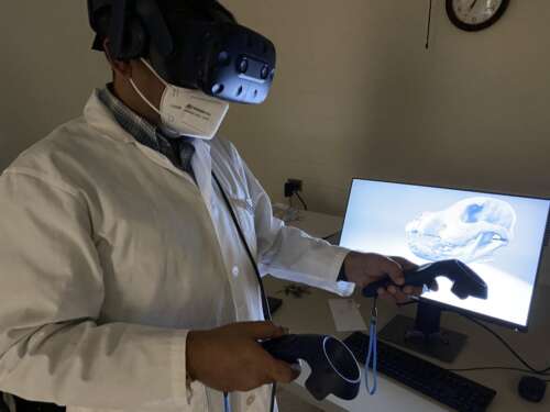
Veterinary students at the University of Guelph will begin using a virtual reality (VR) simulation tool this semester to help learn about dog and cow anatomy.
The technology – believed to be the first use of VR for teaching anatomy at a Canadian veterinary college – allows students to move around virtually inside an animal’s body for a close-up look at organs and tissues.
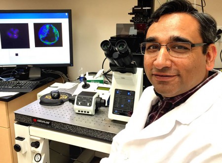
Besides helping to teach anatomy alongside the college’s veterinary anatomy labs, VR tools can help students review material and reduce teaching dependency on preserved specimens, said Dr. Pavneesh Madan, a professor in the Ontario Veterinary College’s (OVC) Department of Biomedical Sciences.
“Such tools have been shown to bring an element of ‘game-ification,’ which students love as they are used to such technology in their daily lives,” he said.
Most important, Madan said, the VR technology will give DVM students a glimpse of the future of veterinary medicine.
“As a university, we are in the profession of training the next generation,” said Madan, who is OVC’s course coordinator for veterinary anatomy. “I think virtual reality becomes a very powerful tool from a futuristic point of view.”
Veterinary anatomy comes to life
Developed in 2018 at the Virginia-Maryland College of Veterinary Medicine, the pertinent software is available to schools under open licence. That means OVC paid only for two VR headsets and associated hardware, now housed in a dedicated room near OVC’s existing anatomy lab.
Madan is using the device this semester to develop protocols for first-year anatomy classes. Students will be able to book time to use the tools to complement their lab training.
Donning a headset, students can “move around” inside the virtual animals to examine and label parts. They can use handheld controls to manipulate the VR model to examine material from any direction, magnify or reduce body parts, and zero in on organs and tissues in three dimensions. Clicking a button allows users to strip away overlying bone to focus on internal organs and systems.
“Anatomy comes to life,” said Madan, who tested the technology along with staff technicians during the past year while setting up the equipment. Referring to VR as a foundation for the “meta-verse,” he added, “You forget the real world, you forget where you are. The tool is like magic, like something I’ve never experienced before.”
The software includes quizzes for students to complete.
Madan said this experiential and immersive learning technology will also help prepare students for future veterinary practice. The VR technology may ultimately be used for planning and simulating surgical operations.
Further in the future, he said, augmented VR may one day enable a practitioner to “visit” patients or attend procedures as a hologram model without leaving the clinic.
“This is a flavour of the times to come.”
OVC is collaborating with Virginia Tech by providing feedback to help the developers hone the technology.
Contact:
Dr. Pavneesh Madan
pmadan@uoguelph.ca