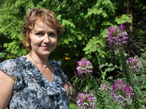
We see plants on a daily basis but we rarely get to see what’s happening inside their cells – until now. “The cells are the basic building blocks of life,” says research associate Nina Barabas, who works in Prof. Jaideep Mathur’s Laboratory of Plant Development and Interactions in the Department of Molecular and Cellular Biology. “We are all made up of cells. Everyone can relate to that.”
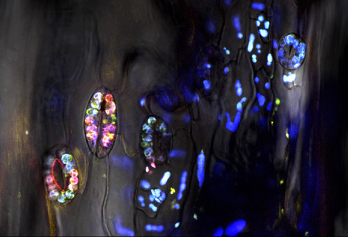
Mathur’s lab uses different types of microscopes to produce stunning images that look more like works of art than plant cells. CellScapes, an exhibition of these images, will be on display Nov. 19 from 3 to 5 p.m. in the Science Complex atrium and from Nov. 20 to 25. in the McLaughlin Library.
The images will also be available to download for free as part of the CellScapes catalogue and will be included in the electronic Atlas of Model Plant Cells. The atlas is geared toward an academic audience, says Barabas, whereas the exhibition is aimed at students of all ages, the general public and anyone who is interested in plants.
Each image in the multimedia atlas is accompanied by a detailed description and contains links to more information and videos. “There are advantages to having an electronic book as a new way of presenting scientific information,” says Barabas. “We’re hoping this electronic resource will reach an international audience.”

The advantages of the e-atlas include interactive images that can be enlarged to show greater detail and snapshots of videos that can be played. Textbooks only contain two-dimensional static images, she adds, whereas the atlas contains three- and four-dimensional images that include depth and time, bringing the images to life. The atlas can also be constantly updated with new content instead of waiting for the next edition of a textbook to be published, which could take years. E-books are also more affordable and portable for students.
The images were made using confocal microscopes, scanning electron microscopes and volume rendering. To make the plant cells glow, coloured fluorescent proteins from jellyfish and coral are added to the cells and a laser beam illuminates the organelles to show how they interact. “All of this is happening at a microscopic level,” says Barabas. “People usually don’t see these minute details even though they are everywhere.” To view the videos, visit www.uoguelph.ca/~jmathur/movies.html.
One of the time-lapse videos shows the growth of an Arabidopsis plant, a common weed. It’s considered a model plant because of its small genome, making it easy for researchers to study. It also has a short lifespan, reaching maturity within six to nine weeks.
In the video, the growing plants appear to be “dancing” to their own rhythm, although there is no wind in the greenhouse and the light source never moves. The dancing plants wouldn’t be noticeable to the naked eye. Another video shows a seedling’s growth as it emerges from a seed. “Most people don’t get to see a germinating seed because things happen underground,” says Barabas.
Her passion for plants is evident in her enthusiasm. Holding up an image of a plant cell printed on a greeting card, she says, “It’s so pretty, I would wear a dress in this print.”
In addition to being a certified aromatherapist and herbalist, Barabas has a PhD in biology from the University of Szeged in Hungary. She also leads wildflower walks in Mississauga, Ont., where she lives, and co-authored a field guide called Wildflowers of Riverwood.
“I just love plants,” she says. “I can’t have enough of them. I like everything about them: their shape and smell and taste and their medicinal properties and all the stories they can tell us.”